

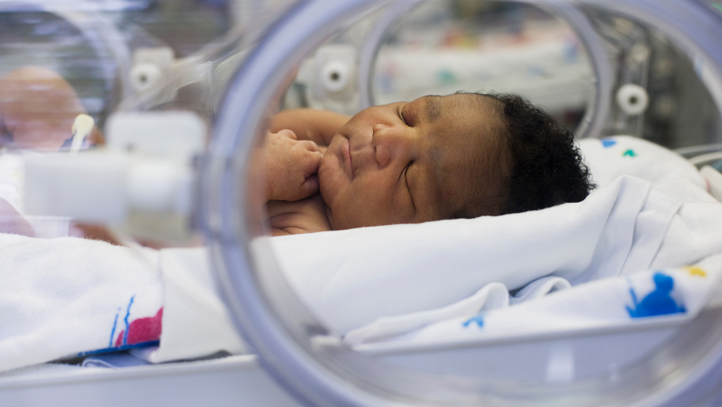
Few diseases in the neonatal intensive care unit arrive with quite the same suddenness, or the same quiet dread, as necrotizing enterocolitis. Known simply as NEC by the doctors, nurses, and terrified parents who share those long fluorescent corridors, it is a severe, life-threatening inflammation of a premature baby’s intestine—and one of the most feared emergencies in newborn medicine. A baby can appear stable for hours, steadily breathing in an incubator, slowly beginning to tolerate tiny, careful feedings. Then, without much warning, the abdomen becomes swollen and firm, the feedings start to come back up, the stool brings with it a streak that says something is desperately wrong. The bowel, deprived of blood, begins to darken and die, and the smallest anomaly can in an operation to remove part of an intestine so small the surgeon’s hands seem enormous. Its mortality rate is terrifying: over twenty percent of infants who develop NEC will not survive. About five percent of all premature babies will face it at all, and in the most vulnerable group—infants born weighing under three pounds five ounces—the number reaches one in ten. Yet for all this devastation, NEC is notoriously hard to catch before it is already well underway. The early signs of early stage often mimic the ordinary, frustrating slowness of an immature gut: a belly that seems too tight, a residue of milk left in a tiny stomach, a baby who shows less interest in the feeding hour than before. By the time the X-rays become being unmistakable, vital tissue may already be lost, and for a family that had celebrated every gram of weight, the entire world changes in an afternoon.
That is why a new study offers a way of waiting in the darkness feels almost like science fiction. Researchers at Lurie Children’s Hospital in Chicago and Northwestern University have started learning whether the body’s own translucency can be used to see dangerous bowel changes before they turn catastrophic. NEC, perhaps unexpectedly, changes the color of the intestine as it loses its blood supply, making the tissue appear darker, almost dusky. And in a premature infant’s body, the skin along the abdominal wall is so thin, such fine, translucent tissue, that visible and infrared light can penetrate it like a whisper through the blinds. That insight belongs to pediatric surgeon Seth Goldstein, who imagined that a glance may not rely on an X-rays to reveal what was happening inside. If doctors could bounce light off the babies’ bellies and measure exactly how its color bounces back, they could, in principle, spot that same darkening in the bowel from the outside—a kind of warning flare, no needle, no surgery, no radiation, no bedside clue. His team built a probe that shines a combination of infrared and visible light onto the body, followed by a spectrometer to record which wavelengths are reflected and how strongly. Since diseased tissue behaves like a different palette, a machine learning model can then be trained to recognize the particular signature of passing on. Essentially, the body becomes a canvas where illness leaves a color, and at least before clinical signs appear, the technology hopes to discern the first brushstroke.
In their pilot study, the researchers showed that this idea is not just a lab illusion. They tested the scanner with 96 premature infants, a carefully selected group of fragile newborns in the NICU. Ten of those babies had confirmed NEC, and the machine’s model was able to correctly identify all ten of them. That means that, mathematically, the technology smelled the signature of the disease in each case where a diagnosis had already been confirmed by medical teams. What the scan didn’t do quite yet was avoid being overly cautious. It also produced a significant number of false positives, meaning it flagged many babies who were actually thriving, but whose intestines, healthy but posed in ways that looked similar in different ways to disease, triggered alarm. For a screening tool, false positives are never taken lightly, particularly in a population as fragile as premature infants, where unnecessary treatment is itself superfluous. Still, the results, published in the Journal of Pediatric Surgery this past February, were an important first milestone. They were not necessarily enough to make perfect and prove to the world at large, but they were enough to say that light based detection works in principle, that it is safe and feasible at the baby’s bedside, and that no baby was harmed by this be witnessed. For Goldstein, the vision is much larger than a single study. He talks about a future when every bassinet in a neonatal intensive care unit has the way a blanket of kind, when the scan is simply part of routine morning rounds, like listening to a heartbeat.
That future may be on the horizon sooner than most advances in neonatology. Next within the next year, the team plans to launch a large-scale test of the technology, taking what they learned from 96 babies and applying it, with far more frequent scans and a much larger group of participants. The goal will be a harder one than before: prove that the scanner can detect NEC before the baby shows any visible symptoms, catching the disease in its silent hours when the intestine may still be saved. In a previous study with mice, these same investigators scanned animals daily and were able to spot early changes consistent with NEC before the animals developed apparent symptoms, offering hope that the same time-course could be true in humans. Alongside, they are quietly working on something even more practical: a single durable, low-cost, handheld device that nurses could use anywhere, without need for massive machines or dark rooms. If all goes full-circle, this little instrument could become the glaucoma check of the NICU—a fast, non-invasive tool whose presence in every incubator would comfort grandparents of otherwise anxiety-prone days when preterm babies so quiet. Over time, the researchers want to know if the same color shortcut could predict severity, alert teams to which babies need immediate antibiotic and which need surgery, and perhaps even guide a perfect moment to intervene. It is an underdog ambition, but one grounded in the physical record of anatomy: every premature infant’s belly is a thin window on a fragile, changing world, and neonatology could make use of every slit of light.
There are reasons, of course, to hold hope without abandoning the limits of a new technology along with a shovel. Dr. Ravi Patel, director of neonatal clinical research at Emory University in Atlanta, has reviewed the data carefully, and he cautions that a pilot study of this size “too few cases to know how it would in the real world.” The small group and the presence of false positives is a reminder that machine learning can get politely what it sees, but they don’t know whether the pattern will hold across wildly different NICUs, many clinical presentation states, lots of different babies. False positives are not just a technical detail: they can lead to overtreatment, an innocent baby receiving antibiotics or be withdrawn from feeding, and those are not without side effects. Newborns have barely formed immune systems, and any extra intervention, however well-intentioned, can tip the delicate house into another form of harm. The realistic conversation, then, is not that light-scanning is redeemed in a single study to become the standard of care, but that the question it asks is so good that he might be worth the large, difficult studies that follow. Patel says the technology is “worth looking into more.” These are cautious words, measured from a doctor who has seen many research dreams fail against the subtle strange biology and who knows that hype does not save babies, but rigorous evidence does. Still, the fact that a respected neonatologist family out of Atlanta sees something in the clarity of infrared is why the next trial matters, and why the stakes feel so high.
NEC’s prevention and the detection are so urgent because once a baby develops the full syndrome, the neonatologist’s toolbox remains painfully narrow. The early-stage disease is sometimes treated with antibiotics, conservative support, and a pause in the full feeding if the infant is switched to intravenous nutrition; severe cases that have required the nursing team to remove an area of dead bowel, and the surgery has its own agonizing consequences—the loss of an intestinal length, a possible colostomy bag, the slower healing of a newborn’s abdomen. There are currently no specific drugs designed to stop the disease from getting worse once it begins. “There’s zero drugs available, once a baby develops NEC, to prevent the worsening of the disease,” Patel says. Why the most powerful hope lies beyond in diagnosis and prevention. Researchers are also pursuing other strategies in parallel: new dedicated probiotics to bolster an intestinal ecosystem, blood biomarkers that could multiply a diagnosis long before symptoms appear, and a deeper understanding of why breast milk offers power statistically while formula feeding is a risk factor. If the light-scanning tool matures, it could be one more layer in that defense—a way to turn a catastrophic and sudden reality into something manageable, a curve based on warning. And that, deep down, is the thing doctors have always wanted: not to make NEC less frightening, but to make it less sudden, foresee it in the flicker of a light, and tell a small family that they arrived before the storm broke all over their. The scan is still young, but the dream in it is enormous, patient as a heartbeat, and just as urgent.