



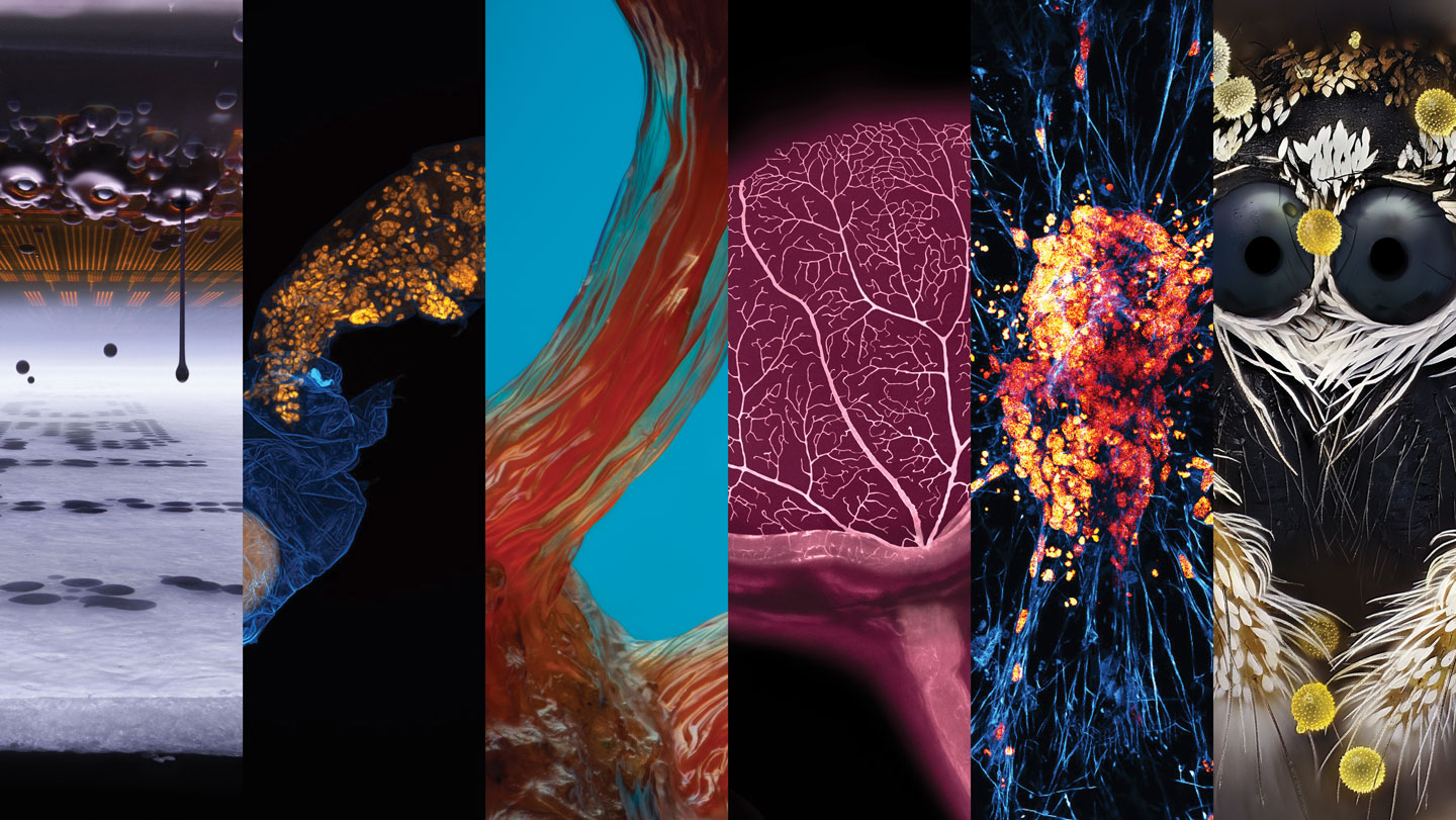
Inside a rebuilt inkjet printer, a tiny universe hangs suspended in midair. Seen through a microscope, individual droplets of ink hover above the page like perfectly spaced beads, revealing the remarkable precision hidden inside one of the most ordinary machines in our homes. This extraordinary image, captured by Ole Bielfeldt, a design student in Cologne, Germany, won first place in the 2026 Nikon Small World photomicrography competition. What makes the image even more remarkable is the story behind it. Bielfeldt’s fascination with the microscopic world began almost by accident during the pandemic. Stuck at home and looking for a way to explore, he pulled an old school microscope out of the attic and started photographing whatever he could find beneath the lens. What began as a quarantine hobby slowly transformed into a serious artistic practice. For the winning photograph, he didn’t simply place a camera against a microscope and hope for the best. He rebuilt an inkjet printer, designing a custom rig that moved the paper instead of the printer’s ink nozzles. By keeping the nozzles stationary and perfectly in focus, he was able to capture the exact moment ink breaks free from its cartridge and begins its journey to the page. The result is an image that makes the familiar strange and the invisible visible, turning something as routine as printing a document into a moment of breathtaking beauty.
The technical challenge of capturing that image was immense. Bielfeldt estimates that the ink droplets were traveling at nearly twenty meters per second, far too fast for even the best slow-motion camera to resolve. To freeze their motion, he worked in complete darkness, illuminating the scene with a single brief flash of light. This technique allowed him to stop time, capturing the droplets exactly as they hung in the air. He instructed the printer to repeatedly spell out the word “macro,” a playful nod to his social media handle, macrofying. Before diving into this project, Bielfeldt admitted that a printer felt “like magic.” He knew it worked, but he never understood how. Photographing its inner workings helped him see the intricate mechanics, the precise timing, and the delicate physics that make printing possible. Now, he says, he no longer takes the magic printers spit out for granted. “It’s so, so advanced technically, but at the same time it’s so, so common,” he explained. That sense of wonder is at the heart of all the images recognized in this year’s competition, whose winners were announced on October 21. Across eighty-six photographs, scientists and artists alike captured the hidden beauty of the natural world, from the deepest structures of living tissue to the smallest creatures that share our planet.
Among the standout images was a stunning portrait of a pollen-dusted jumping spider, captured by John-Oliver Dum. At first glance, the spider looks almost mythical, its body covered in bristling hairs and its eight eyes gleaming, details that are easy to miss with the naked eye. Dum’s fascination with spiders began, surprisingly, as a childhood fear. He suffered a particularly painful arachnid bite when he was young, and for years spiders were something to avoid rather than admire. That changed when he became inspired to study the movements of a curly hair tarantula, and he soon grew passionate about capturing their beauty. His photographs require extraordinary patience and preparation, sometimes beginning years before they are submitted to a competition. His wife grows plants specifically selected for their pollen, which Dum then uses in his images. He described the biggest challenge as finding a spider like this one on the right plants, with the right pollen attached, at exactly the right time of year and at exactly the right moment. It meant understanding not only the spider’s behavior, but also the surrounding insect and plant life, and their seasonal rhythms. His second-place image is a reminder that even creatures we fear can be beautiful when we take the time to truly see them. Dum hopes the photograph encourages viewers to look more closely at the world around them. “We first have to see the beauty of this largely unknown world before we can truly understand what we may be destroying,” he said.
Another image that captured the imagination was a cross section of a mouse eye, crisscrossed with fractal patterns of blood vessels and tethered by the optic nerve. At first glance, it looks more like an exotic leaf or flower than part of an animal, but it represents something deeply scientific. Cell biologists Sinem Karaman and Satu Paavonsalo, at the University of Helsinki, specialize in visualizing vascular networks to understand how blood vessels work within tissues. Their research could even point to mechanisms underlying how these systems stop functioning properly in conditions like obesity and diabetes. For this image, they set out to capture a mouse eye in three dimensions, a departure from the flattened retinas typically used in their research. The mounting process was meticulous and required several attempts before Paavonsalo was satisfied with the arrangement. Even then, she couldn’t be certain the image would turn out correctly. She had to hope that the sample was properly positioned and that the staining was detected throughout the entire sample. That uncertainty made the moment of seeing the final image even more exciting. The photograph earned nineteenth place in the competition and stands as a reminder that even familiar parts of the body, like an eye, contain worlds of complexity yet to be explored. Around the same time, another remarkable creature caught the judges’ attention: a tardigrade, also known as a water bear, captured by evolutionary developmental biologist Gonzalo Quiroga-Artigas. In this image, a tardigrade emerges from its shed cuticle, and behind it lies a surprising treasure: an egg left behind to be protected by its mother’s discarded skin. Tardigrades are among the toughest creatures on Earth, capable of surviving space by entering a state of suspended animation. This particular species, Hypsibius exemplaris, is only about two and a half times as wide as a strand of hair. Quiroga-Artigas captured the image during his studies at the University of Montpellier in France. He explained that, in the tardigrade life cycle, egg laying is closely synchronized with molting, and the moment one sheds its old skin happens so quickly that it is easy to miss. Snapping the image required painstaking mounting and imaging of many specimens, but he chose this one, which won twelfth place, because the egg looks almost like a precious gem being left behind by its mother.
A slime mold photographed in the heart of Brooklyn, New York, also drew widespread attention. Eric Epstein found the mold, a species of Hemitrichia, sprouting from a decaying log in Prospect Park. The image highlights the mold’s gelatinous fruiting bodies, delicate structures that release spores as part of reproduction. To bring every detail into sharp focus, Epstein stacked two hundred and forty images, a technique that allows photographers to create a single image with incredible depth. His process began with careful searching through local parks, cutting pieces of waterlogged wood free without disturbing his tiny subjects. In their liquid state, he said, any touch would demolish the fruiting bodies, so extreme care was required. Slime molds are more fascinating than most people realize. One species closely mirrors the cosmic web of gas and dark matter that connects galaxies, helping scientists map the universe more accurately. Another species has fascinated researchers because it can pack away a store of bacteria to feed the next generation when it is near death. Epstein hopes his photograph hints at the vast array of activity happening beyond our unaided perception. The image won sixth place in the competition. Also honored was a striking image of a neuron by undergraduate student Faith Fernandes and structural cell biologist James Glover at the University of California, Berkeley. The image shows clusters of nuclei that look like flashpoints in an intricate web, commemorating the team’s first attempt at crafting neurons from a particular kind of stem cell. Fernandes said she was so instantly enamored with the piece that she made it her computer wallpaper as soon as it was taken. The brilliant colors depict human induced pluripotent stem cells, which have the remarkable ability to become any type of cell in the body, that have developed into neurons. In the image, neurotubules appear blue, while orange indicates where DNA is located. These stem cells are created by reprogramming adult cells back to an earlier stage, before they were locked into becoming a specific cell type. Researchers study them for their potential in regenerative medicine, including repairing damaged tissue and addressing diseases like Parkinson’s. Already, scientists have used these cells to regrow eye tissue, address premature aging in mouse studies, and grow organs in the lab. The image earned an Image of Distinction title, and for Fernandes, it represents hope for new ways to understand and treat neurodegeneration.
Taken together, these images do more than showcase scientific achievement; they invite us to pause and marvel at the world we usually overlook. Each one required not only technical skill but also an artist’s eye and a scientist’s patience. From the ink droplets inside a common printer to a spider dusted with pollen, from a mouse eye’s hidden blood vessels to a tardigrade’s jewel-like egg, from the delicate fruiting bodies of a slime mold to a neuron blazing like a galaxy, these photographs reveal that beauty and complexity exist everywhere, even in the smallest corners of life. Bielfeldt’s journey is particularly human: he started with an old microscope and a quarantine hobby, and found himself transformed by curiosity. He no longer sees printers as magic because he now understands how they work, but perhaps that understanding has revealed an even deeper magic. The competition celebrates this kind of discovery, reminding us that wonder is not limited to distant galaxies or deep oceans. It is present in the objects we use every day, in the creatures that share our neighborhoods, and in the cells that make up our own bodies. As John-Oliver Dum suggested, we first have to see the beauty of this largely unknown world before we can truly understand what we may be destroying. These images give us that chance. They ask us to look closer, to slow down, and to remember that the most ordinary things, when seen through the right lens, can become extraordinary.