







For decades, a silent and deeply terrifying specter has hovered over the worlds of contact sports, military combat, and physical trauma: Chronic Traumatic Encephalopathy, commonly known as CTE. This progressive, degenerative brain disease, triggered by repeated sub-concussive and concussive blows to the head, has quietly shattered the lives of countless athletes, military veterans, and their loved ones, leaving behind a painful trail of cognitive decline, severe mood swings, erratic behavioral changes, and tragic instances of suicide. Yet, despite the growing public awareness and the profound human tragedy surrounding this condition, a frustrating and agonizing barrier has remained stubbornly unbroken: CTE can currently only be diagnosed after death. To confirm a diagnosis, pathologists must meticulously slice and examine the physical brain tissue of the deceased, leaving living individuals trapped in a state of tormenting psychological limbo, experiencing a terrifying decline in their mental faculties without ever receiving a definitive medical confirmation of what is happening inside their minds. This lack of diagnostic capability not only deprives patients of closure but also paralyzes scientific efforts to develop effective treatments, as researchers cannot treat a disease they cannot accurately identify in the living. However, a momentous breakthrough presented at the Alzheimer’s Association International Conference in London has offered a powerful, long-awaited glimmer of hope to those living under this shadow. Groundbreaking preliminary data suggests that a specific protein biomarker, originally developed to identify Alzheimer’s disease, could pave the way for the world’s very first diagnostic tool for CTE in living patients. By detecting and measuring a protein called eMTBR-tau243, scientists believe they may have unlocked a biological window into the living brain, potentially revolutionizing how we understand, identify, and support those who have dedicated their lives to high-impact activities, shifting the narrative of CTE from a tragic posthumous revelation to an active, treatable medical reality.
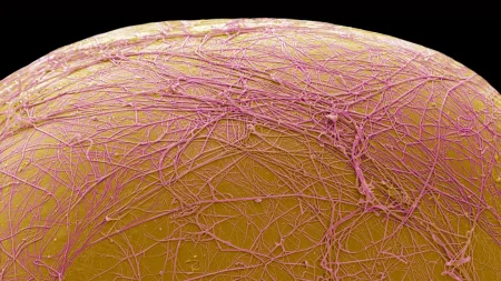
To appreciate the magnitude of this discovery, it is essential to understand the microscopic devastation that occurs within a brain affected by CTE, which is classified scientifically as a tauopathy. In a healthy, functioning brain, a protein known as tau plays a vital, supportive role, serving as a sort of cellular logistics manager or structural scaffolding system that stabilizes microscopic pathways called microtubules. These microtubules act as internal highways, transporting essential nutrients, signaling molecules, and biological materials across brain cells to keep the mind sharp, stable, and healthy. However, when the brain is subjected to repeated, violent physical forces—such as the routine tackles in American football, the head impacts in boxing, or blast injuries in combat—this delicate cellular infrastructure is violently disrupted. The tau proteins break away from their structural posts, undergoing abnormal chemical changes that cause them to warp, misfold, and clump together into toxic, tangled deposits. These sticky tau tangles gradually spread throughout the brain, choking off healthy neurons and cutting off their communication pathways, which ultimately leads to the progressive death of brain tissue and the shrinkage of vital brain regions. This cellular destruction manifests outwardly as the severe cognitive, emotional, and physical impairments characteristic of the disease. While a 2018 study suggested that CTE-like damage was present in roughly one out of every 164 donated brains from the general public, the statistics are shockingly higher for those repeatedly exposed to head trauma. In 2023, the Boston University CTE Center published a staggering report revealing that CTE pathology was present in more than 90 percent of the brains studied from a sample of 376 former National Football League (NFL) players, underscoring the urgent, systemic need for an early-warning diagnostic system that can identify this microscopic damage before the behavioral symptoms turn fatal.
As is so often the case in the history of medicine, this profound scientific breakthrough was not the result of a direct search for a CTE diagnostic, but rather a remarkable stroke of scientific serendipity. The journey began at Washington University in St. Louis, where neuroscientist Chihiro Sato and her colleague, Kanta Horie, were focused on a completely different neurological challenge: developing a highly sensitive and specific diagnostic test for Alzheimer’s disease. Like CTE, Alzheimer’s is characterized by the accumulation of destructive tau tangles, although these tangles accumulate in different regions of the brain and follow a distinct pathological pattern. Working in close collaboration with the pharmaceutical company Eisai Inc., which is actively developing therapeutic treatments targeting tau dysfunction, the researchers were targeting a specific fragment of the tau protein known as eMTBR-tau243, hoping to prove that its elevation in bodily fluids was entirely unique to Alzheimer’s and would not trigger false positives in patients suffering from other neurodegenerative conditions, such as progressive supranuclear palsy. To test this specificity and ensure their marker was a reliable diagnostic tool, Horie analyzed cerebrospinal fluid samples taken from a diverse cohort of 112 patients suffering from eight different brain diseases. While the test successfully proved highly specific to Alzheimer’s across the board, the researchers encountered a “very surprising” outlier that defied all of their expectations: a single patient who did not have Alzheimer’s but nevertheless exhibited extremely elevated levels of eMTBR-tau243. Upon closer investigation, the researchers discovered that this patient suffered from advanced, severe CTE, leading them to the sudden, exciting realization that the structural characteristics of tau clumps in both Alzheimer’s and CTE are similar enough that the same molecular test could detect them both, opening up an entirely unplanned path toward a living CTE diagnostic.
Eager to explore this unexpected connection, Sato, Horie, and their research team quickly pivoted, partnering with expert researchers at the University of California, San Francisco to gather a larger and more targeted set of patient samples. Because CTE can only be definitively diagnosed after death, the team had to work backward, obtaining precious samples of blood and cerebrospinal fluid that had been collected from individuals while they were still alive, and who had subsequently donated their brains to science upon their passing. This unique and invaluable dataset included blood samples from 11 deceased individuals whose post-mortem brain autopsies had officially confirmed the presence of varying stages of CTE pathology. When the scientists analyzed these blood samples, the results were nothing short of extraordinary: they discovered that the eMTBR-tau243 signal in the blood did not just indicate the presence of the disease, but actually increased in a clear, stage-specific manner that mirrored the physical progression of the brain damage. Donors who had passed away with mild, Stage I CTE exhibited significantly lower levels of the protein in their blood compared to those who had died with advanced, Stage IV disease, while all of the CTE patients showed levels that were significantly higher than those found in healthy, age-matched control subjects. Even though the team was only able to acquire two brains with moderate Stage III disease, the overall trend clearly suggested that their biomarker levels would sit perfectly on the spectrum between the early and late stages, providing compelling evidence that a simple, minimally invasive blood test could one day determine not just if a person has CTE, but how far the disease has progressed.
This biological breakthrough comes at a critical juncture in the field of neurology, where previous efforts to diagnose CTE through behavioral symptoms and cognitive assessments alone have repeatedly hit frustrating dead ends. In 2019, a specialized panel of international experts attempted to address this clinical diagnostic void by developing a detailed diagnostic checklist, compiling a set of likely symptoms, behavioral patterns, and history of repetitive head impacts that might allow clinicians to identify living patients with probable CTE. However, relying on subjective behavioral assessments has proven to be an unreliable and deeply flawed approach, as human behavior is far too complex, varied, and easily influenced by outside factors to be categorized on a standardized form. A rigorous study published in May by neurosurgeon John Arena of the University of Pennsylvania and his colleagues revealed a sobering reality: there was almost no statistically significant correlation between patients who scored highly on this clinical checklist during their lives and those whose post-mortem autopsies actually confirmed the physical presence of CTE pathology in their brains. Because the prominent symptoms of CTE—including severe depression, intense anxiety, irritability, memory lapses, and executive dysfunction—overlap so heavily with other common psychiatric disorders, post-traumatic stress disorder (PTSD), and the natural process of aging, clinical symptom checklists on their own are simply insufficient to provide a definitive diagnosis. As Arena strongly emphasizes, these findings underscore why the medical community cannot rely solely on observation, highlighting the critical and desperate need for validated, objective biological biomarkers in blood or spinal fluid to supplement the clinical picture, as a concrete, laboratory-verified blood test would eliminate subjective guesswork, prevent devastating misdiagnoses, and finally provide patients with the indisputable, scientific clarity they deserve regarding the physical health of their brains.
Looking toward the future, the implications of a validated, living biomarker for CTE are profound, offering a pathway toward a new era of medical hope, therapeutic innovation, and psychological relief. For the millions of individuals who have sustained repeated head injuries—from professional athletes and youth sports participants to military veterans and survivors of domestic abuse—the ability to receive a definitive diagnosis in life would lift an immense emotional burden, validating their internal struggles and allowing them to make informed decisions about their careers, lifestyles, and long-term healthcare. Furthermore, as neurosurgeon John Arena points out, the development of a reliable diagnostic blood test is the essential, missing key required to unlock clinical trials for potential CTE treatments. Currently, pharmaceutical companies are severely hindered in their ability to develop therapies because they cannot accurately identify living trial participants who actually have the disease, nor can they measure whether an experimental drug is successfully halting the progression of tau tangles in real-time. By utilizing eMTBR-tau243 as a reliable benchmark, researchers could finally enroll confirmed CTE patients in rigorous clinical trials, monitor the efficacy of new medications through routine blood draws, and accelerate the development of targeted therapies designed to stop the disease in its tracks. While Chihiro Sato and her colleagues caution that these preliminary findings must still be validated in much larger, more diverse patient datasets, this pioneering research represents a monumental leap forward, transforming our approach to brain trauma and offering a bright beacon of hope that CTE will one day be a treatable, preventable condition rather than a silent, posthumous tragedy.