



For decades, the iconic double helix of DNA has been celebrated as the blueprint of life, a miraculously elegant structure that encodes the instructions for every living organism. Yet, this iconic image often conceals a perplexing biological paradox that has stumped scientists for years. DNA is a heavily negatively charged molecule, a property that should, according to the fundamental laws of physics, cause any two strands to repel each other with immense force, like two powerful magnets with matching poles held close together. But inside our cells, DNA is packed impossibly tight—roughly two meters of it is folded into a nucleus just six microns wide. For years, the scientific community assumed that only bulky proteins like histones could overcome this electrostatic repulsion, acting as spools to wind the DNA around. However, a groundbreaking new study, combining an extraordinary instrument that resembles a vintage record player with massive supercomputer simulations, has cracked this long-standing mystery wide open. The answer, as it turns out, lies in an invisible cast of characters: tiny metal ions. These metallic particles, acting like microscopic rivets or molecular glue, can spontaneously slip between the negatively charged backbones of two DNA strands, physically bridging them together and overriding the natural repulsion. This discovery not only solves a fundamental question in biophysics but reshapes our entire understanding of how genetic material is structured, stored, and manipulated in living systems.
The electrostatic paradox that plagued scientists is rooted in the very chemical structure of the DNA molecule. Each strand is composed of a sugar-phosphate backbone, and each phosphate group carries a net negative charge. In a physiological solution, this creates a thick, repulsive cloud of electron density that surrounds the helix. In the 1960s and 1970s, researchers studying DNA condensation discovered that they could force naked DNA strands to bundle together by adding certain salts to the solution. The logic was simple: the positively charged ions in the salt would cluster around the negative DNA, effectively “shielding” the charge and allowing the strands to approach. Yet, this theory only worked up to a point. Monovalent ions like sodium could disperse the charge, but they could never bring the strands into tight contact; the residual repulsion was simply too strong. The mystery deepened when they found that divalent ions—those carrying two positive charges, like magnesium, zinc, and cobalt—could achieve this tight packing. But how? Standard electrostatic theory predicted that even divalent ions would rapidly diffuse away from the space between two strands, unable to stay put long enough to provide a stable adhesive force. This was the “ion condensation” contradiction: the ions should neutralize the repulsion, but shouldn’t be able to bridge the strands, because the thermal motion of the molecules should constantly push them out of the gap. The conflict between these theoretical models and the clear experimental evidence that DNA strands could spontaneously form highly ordered, crystalline bundles left the field without a coherent explanation for decades.
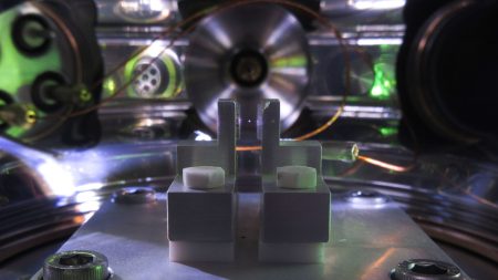
The key to finally observing this elusive phenomenon came from an unlikely source: an instrument inspired by the mechanics of a vinyl record turntable. This is the Atomic Force Microscope (AFM), a remarkable device that operates on a completely different principle than traditional electron microscopes. Electron microscopes require a vacuum and bombard the sample with beams of electrons, which destroys the delicate, hydrated environment in which DNA naturally exists. The AFM, in contrast, works in a liquid buffer, mimicking the conditions inside a living cell. It operates by tracing a sharp, microscopic “needle” attached to a flexible cantilever across the surface of the sample. As the needle moves, it physically “feels” the bumps and valleys of the molecules, and a laser detects the minute bending of the cantilever to create a three-dimensional topographical map. By carefully controlling the speed and force, the researchers were able to lay down dilute solutions of DNA onto a flat mica surface, inject solutions containing various metal ions, and then use this “record player” to scan across the surface. What they saw was astonishing: individual DNA strands, effectively naked without any proteins, were aligning themselves in parallel, tightly packed bundles. The distance between the strands was less than one nanometer, far too close for mere electrostatic shielding. It was direct, physical evidence that something invisible was physically locking them together.
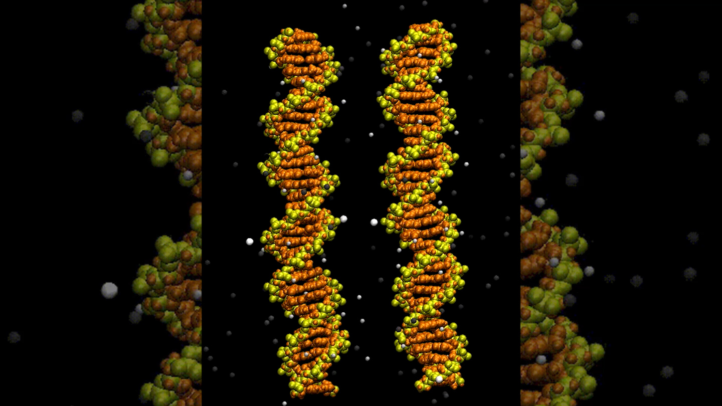
To interpret these microscopic images, the team turned to the digital realm, building intricate computer simulations that modeled the behavior of every single atom in the system. Think of this as a digital sandbox where they placed two DNA helices inside a virtual cube filled with water molecules and various metal ions. Using the laws of physics—mapping the attractions and repulsions of every electron—the supercomputers calculated how each ion moved over time, both in the bulk solution and between the strands. The results defied conventional wisdom. Instead of the ions merely floating around, the simulations revealed a dynamic yet persistent phenomenon. When a divalent metal ion (such as zinc) approached the gap between two adjacent DNA strands, it would get momentarily trapped. The electrostatic attraction from the two opposing phosphate backbones would pull the ion into a tight coordination pocket. But instead of bouncing right out, the ion would form multiple bonds simultaneously—linking one oxygen atom from one DNA strand to an oxygen atom from the other strand. This created a literal “salt bridge” or “ion bridge.” The simulation showed that while a single bridge was fleeting and would break within nanoseconds, the sheer statistical probability of multiple ions forming these bridges at the same time, across the entire length of the DNA, generated a powerful, collective adhesive force that was robust enough to overcome the electrostatic repulsion permanently.
The fusion of the experimental microscope images and the computational simulations revealed the “eureka” moment. The record-player microscope proved that DNA strands could get physically close; the simulations explained how the metal ions executed this feat. The magnesium or zinc ions, due to their small size and high charge density, are attracted to the negative phosphate groups, but crucially, they act as powerful coordinating agents. They strip away a layer of water molecules and snuggle directly into the grooves between the DNA backbones, tightly binding to the phosphate oxygen atoms of both strands. This binding is not a static, permanent weld but a delicate dance of rapid association and dissociation. However, the avalanche of these transient connections acts like the subtle, relentless pressure of a thousand tiny hands, pulling the DNA strands together and holding them locked in place. This mechanism easily explains why calcium, zinc, and cobalt are so effective at condensing DNA in test tubes. More importantly, it reveals that DNA does not need a protein spool to pack itself tightly inside a virus or during certain stages of cell division. The cell itself can utilize the very ions floating in its cytoplasm to orchestrate packaging. This solves the riddle of how certain viruses, which lack histones, can cram their enormous genomes into tiny protein shells with such astronomical density.
The implications of this discovery ripple far beyond a textbook update on molecular physics. It provides a new, foundational framework for understanding the fundamental mechanics of life. For nanotechnologists, this opens up a robust new tool for building “DNA origami” or designing synthetic genetic circuits, because the precise control of ion concentration could be used as a switch to control how tightly DNA folds and unfolds. In medicine, this could transform gene therapy, where the primary hurdle is delivering fragile DNA into cells. If we can now tune the metal ion environment to create perfectly compact, stable “DNA packages,” we could improve the efficiency of delivering therapeutic genes to treat genetic diseases. Furthermore, this research offers a glimpse into the beautiful complexity of nature’s engineering. It shows that the biological world is not just a story of giant, complex proteins, but also of the elegant choreography of the smallest particles. Just as a record player translates tiny grooves into beautiful music, this unique microscope has translated the invisible physical interactions of atomic matter into a clear melody of understanding. The mystery of how DNA overcomes its own repulsion is solved, but it also serves as a humble reminder that the secrets of biology often hide in plain sight, resolvable only when we combine the sharpest experimental eyes with the profound predictive power of computational theory. It is a triumph of interdisciplinary science, and a testament to human curiosity that even the most fundamental, taken-for-granted aspects of life—the very substance that makes us who we are—still hold the power to surprise and inspire us.